
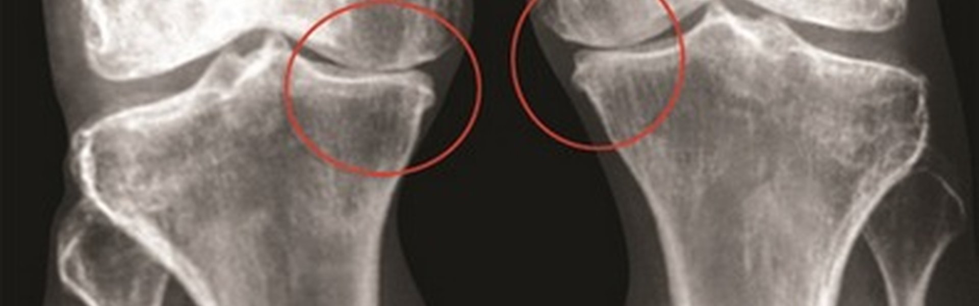
Osteoarthritis treatment
Our company supports the preparation of patients for arthro-medullary shunting — a joint-preserving treatment approach that, in some cases, may help delay or avoid joint replacement.
Arthro-medullary shunting uses the patient’s own biological resources to support treatment of the affected joint. The procedure is aimed at reducing pain, improving the conditions for joint cartilage function, and restoring joint mobility. Following surgery, patients may require fewer analgesics and other symptomatic treatments.
The primary goal of the procedure is to preserve the patient’s natural joint and maintain its function for as long as possible, thereby delaying the need for joint replacement.
How Osteoarthritis Develops
Degenerative joint diseases are serious disorders of the musculoskeletal system. As osteoarthritis progresses, the properties of synovial fluid and joint lubrication may deteriorate, inflammatory processes may intensify, and cartilage damage may develop. Changes also affect the subchondral bone located directly beneath the articular cartilage and may be accompanied by increased intraosseous pressure.
These changes can lead to pain, stiffness, and restricted joint movement, significantly affecting patients’ quality of life.
As the disease progresses, joint replacement may become necessary in some cases. Arthro-medullary shunting was developed as a joint-preserving approach aimed at maintaining the affected joint. The technique is primarily used for knee disorders and is also applied in the treatment of ankle joint pathology.
How the Arthro-Medullary Shunt Works
The arthro-medullary shunt creates a localized channel between the bone marrow cavity and the joint cavity. Through this channel, the patient’s own bone marrow fat enters the joint, where it may contribute to joint lubrication and reduce friction between the articular surfaces.
Precursor cells from the bone marrow may also enter the joint together with the bone marrow fat and may contribute to regenerative processes in damaged cartilage.
Importantly, the procedure involves the bone marrow located within the bone, not the brain, spinal cord, or cerebrospinal fluid. The shunt is placed directly into the bone in the area of the affected joint, creating a controlled pathway between the bone marrow cavity and the joint cavity.
Implant Design and Placement
The arthro-medullary shunt is made of a titanium alloy and consists of a hollow tube with a diameter of 8 mm and a length of 60 to 120 mm. During a minimally invasive surgical procedure, the shunt is placed into the femoral condyle. A threaded section provides secure fixation within the bone, while lateral openings allow bone marrow fat to enter the inner channel of the shunt and then the joint cavity.
The implant is placed directly in the area of the affected joint and does not involve the spine, spinal cord, or other structures of the central nervous system. The minimally invasive placement technique is designed to minimize surgical trauma and preserve the surrounding tissues.
The safety of the procedure depends on appropriate patient selection, proper surgical technique, and postoperative monitoring. A preoperative assessment is performed to evaluate the condition of the joint and determine whether the procedure is appropriate.
For more information, please contact us.